Cotton Wool Bodies . They have been described in many. Usually do not produce vision loss unless large or near fovea. Cotton wool spots (cwss) are localised accumulations of axoplasmic debris within adjacent bundles of unmyelinated ganglion cell. While the spots themselves don’t. Cotton wool spots are opaque fluffy white patches on the retina of the eye that are considered an abnormal finding during a funduscopic exam. Cotton wool spots (cws) are fluffy white or yellow spots that can appear on the retina. White spots on retinal surface caused by microinfarction.
from www.allaboutvision.com
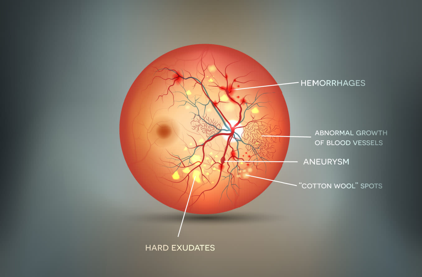
Cotton wool spots (cws) are fluffy white or yellow spots that can appear on the retina. White spots on retinal surface caused by microinfarction. They have been described in many. While the spots themselves don’t. Cotton wool spots (cwss) are localised accumulations of axoplasmic debris within adjacent bundles of unmyelinated ganglion cell. Usually do not produce vision loss unless large or near fovea. Cotton wool spots are opaque fluffy white patches on the retina of the eye that are considered an abnormal finding during a funduscopic exam.
Cotton Wool Spots Causes and Symptoms
Cotton Wool Bodies White spots on retinal surface caused by microinfarction. White spots on retinal surface caused by microinfarction. Cotton wool spots are opaque fluffy white patches on the retina of the eye that are considered an abnormal finding during a funduscopic exam. While the spots themselves don’t. Cotton wool spots (cws) are fluffy white or yellow spots that can appear on the retina. Cotton wool spots (cwss) are localised accumulations of axoplasmic debris within adjacent bundles of unmyelinated ganglion cell. They have been described in many. Usually do not produce vision loss unless large or near fovea.
From www.alamy.com
Heap of soft cotton wool on white background Stock Photo Alamy Cotton Wool Bodies Cotton wool spots (cwss) are localised accumulations of axoplasmic debris within adjacent bundles of unmyelinated ganglion cell. White spots on retinal surface caused by microinfarction. Usually do not produce vision loss unless large or near fovea. While the spots themselves don’t. Cotton wool spots are opaque fluffy white patches on the retina of the eye that are considered an abnormal. Cotton Wool Bodies.
From ar.inspiredpencil.com
Grey Cotton Wool Spots Cotton Wool Bodies They have been described in many. Usually do not produce vision loss unless large or near fovea. White spots on retinal surface caused by microinfarction. Cotton wool spots (cws) are fluffy white or yellow spots that can appear on the retina. While the spots themselves don’t. Cotton wool spots are opaque fluffy white patches on the retina of the eye. Cotton Wool Bodies.
From www.allaboutvision.com
Cotton Wool Spots Causes and Symptoms Cotton Wool Bodies Cotton wool spots (cwss) are localised accumulations of axoplasmic debris within adjacent bundles of unmyelinated ganglion cell. While the spots themselves don’t. White spots on retinal surface caused by microinfarction. Cotton wool spots (cws) are fluffy white or yellow spots that can appear on the retina. Cotton wool spots are opaque fluffy white patches on the retina of the eye. Cotton Wool Bodies.
From livewell-group.com
Cotton Wool Livewell Group Cotton Wool Bodies While the spots themselves don’t. White spots on retinal surface caused by microinfarction. Cotton wool spots are opaque fluffy white patches on the retina of the eye that are considered an abnormal finding during a funduscopic exam. Cotton wool spots (cws) are fluffy white or yellow spots that can appear on the retina. They have been described in many. Usually. Cotton Wool Bodies.
From facemask2020.en.made-in-china.com
100 Cotton Wool Surgical Medical Use 500g China Cotton Wool and Cotton Wool Bodies Usually do not produce vision loss unless large or near fovea. Cotton wool spots (cws) are fluffy white or yellow spots that can appear on the retina. While the spots themselves don’t. Cotton wool spots are opaque fluffy white patches on the retina of the eye that are considered an abnormal finding during a funduscopic exam. Cotton wool spots (cwss). Cotton Wool Bodies.
From www.alamy.com
Ball of clean cotton wool isolated on white Stock Photo Alamy Cotton Wool Bodies Cotton wool spots are opaque fluffy white patches on the retina of the eye that are considered an abnormal finding during a funduscopic exam. They have been described in many. Cotton wool spots (cws) are fluffy white or yellow spots that can appear on the retina. While the spots themselves don’t. Cotton wool spots (cwss) are localised accumulations of axoplasmic. Cotton Wool Bodies.
From www.pinterest.co.uk
cotton wool spots vs hard exudates Google Search Optometry, Eye Cotton Wool Bodies White spots on retinal surface caused by microinfarction. While the spots themselves don’t. Usually do not produce vision loss unless large or near fovea. Cotton wool spots are opaque fluffy white patches on the retina of the eye that are considered an abnormal finding during a funduscopic exam. Cotton wool spots (cws) are fluffy white or yellow spots that can. Cotton Wool Bodies.
From www.researchgate.net
Solitary cottonwool spot in the right eye ofa patient with PGL who Cotton Wool Bodies While the spots themselves don’t. Cotton wool spots are opaque fluffy white patches on the retina of the eye that are considered an abnormal finding during a funduscopic exam. Usually do not produce vision loss unless large or near fovea. They have been described in many. Cotton wool spots (cws) are fluffy white or yellow spots that can appear on. Cotton Wool Bodies.
From www.semanticscholar.org
Figure 1 from Detection Of Cotton Wool Spots In Retinopathy Images A Cotton Wool Bodies Cotton wool spots (cwss) are localised accumulations of axoplasmic debris within adjacent bundles of unmyelinated ganglion cell. They have been described in many. While the spots themselves don’t. White spots on retinal surface caused by microinfarction. Usually do not produce vision loss unless large or near fovea. Cotton wool spots (cws) are fluffy white or yellow spots that can appear. Cotton Wool Bodies.
From www.youtube.com
12 COTTON WOOL BALLS REMOVED FROM PATIENTS EARS EP178 YouTube Cotton Wool Bodies While the spots themselves don’t. Cotton wool spots (cwss) are localised accumulations of axoplasmic debris within adjacent bundles of unmyelinated ganglion cell. Cotton wool spots are opaque fluffy white patches on the retina of the eye that are considered an abnormal finding during a funduscopic exam. Cotton wool spots (cws) are fluffy white or yellow spots that can appear on. Cotton Wool Bodies.
From mamaowl.net
Organic Merino Wool, Cotton & Silk Body Pale Pink MamaOwl Cotton Wool Bodies They have been described in many. Cotton wool spots are opaque fluffy white patches on the retina of the eye that are considered an abnormal finding during a funduscopic exam. While the spots themselves don’t. Cotton wool spots (cwss) are localised accumulations of axoplasmic debris within adjacent bundles of unmyelinated ganglion cell. White spots on retinal surface caused by microinfarction.. Cotton Wool Bodies.
From jamanetwork.com
CottonWool Spots and Retinal Hemorrhages Clinical Pharmacy and Cotton Wool Bodies While the spots themselves don’t. Usually do not produce vision loss unless large or near fovea. Cotton wool spots (cwss) are localised accumulations of axoplasmic debris within adjacent bundles of unmyelinated ganglion cell. White spots on retinal surface caused by microinfarction. Cotton wool spots (cws) are fluffy white or yellow spots that can appear on the retina. They have been. Cotton Wool Bodies.
From www.youtube.com
COTTON WOOL SPOTS EXPLAINED ! YouTube Cotton Wool Bodies Cotton wool spots (cws) are fluffy white or yellow spots that can appear on the retina. They have been described in many. White spots on retinal surface caused by microinfarction. While the spots themselves don’t. Cotton wool spots are opaque fluffy white patches on the retina of the eye that are considered an abnormal finding during a funduscopic exam. Cotton. Cotton Wool Bodies.
From webeye.ophth.uiowa.edu
Cotton wool spots. COMS Grading Scheme Cotton Wool Bodies While the spots themselves don’t. Cotton wool spots (cws) are fluffy white or yellow spots that can appear on the retina. Usually do not produce vision loss unless large or near fovea. Cotton wool spots are opaque fluffy white patches on the retina of the eye that are considered an abnormal finding during a funduscopic exam. They have been described. Cotton Wool Bodies.
From bjo.bmj.com
Why cotton wool spots should not be regarded as retinal nerve fibre Cotton Wool Bodies They have been described in many. Usually do not produce vision loss unless large or near fovea. White spots on retinal surface caused by microinfarction. While the spots themselves don’t. Cotton wool spots (cwss) are localised accumulations of axoplasmic debris within adjacent bundles of unmyelinated ganglion cell. Cotton wool spots are opaque fluffy white patches on the retina of the. Cotton Wool Bodies.
From www.first-aid-online.co.uk
Cotton Wool 500g Cotton Wool Bodies Usually do not produce vision loss unless large or near fovea. While the spots themselves don’t. Cotton wool spots are opaque fluffy white patches on the retina of the eye that are considered an abnormal finding during a funduscopic exam. They have been described in many. White spots on retinal surface caused by microinfarction. Cotton wool spots (cwss) are localised. Cotton Wool Bodies.
From clinicaloptometry.scholasticahq.com
Cotton Wool Spots in a Patient with COVID19 Published in CRO Cotton Wool Bodies Usually do not produce vision loss unless large or near fovea. They have been described in many. Cotton wool spots (cwss) are localised accumulations of axoplasmic debris within adjacent bundles of unmyelinated ganglion cell. Cotton wool spots (cws) are fluffy white or yellow spots that can appear on the retina. White spots on retinal surface caused by microinfarction. Cotton wool. Cotton Wool Bodies.
From glenmia.com.au
Cotton Wool roll Glen Mia Saddlery Cotton Wool Bodies They have been described in many. While the spots themselves don’t. Cotton wool spots (cwss) are localised accumulations of axoplasmic debris within adjacent bundles of unmyelinated ganglion cell. Usually do not produce vision loss unless large or near fovea. White spots on retinal surface caused by microinfarction. Cotton wool spots (cws) are fluffy white or yellow spots that can appear. Cotton Wool Bodies.